
Horse science
Parts of the horse's hoof: the frog and the wall
· 4 min read
Like a human fingernail, the hoof wall has no nerves or blood vessels and grows about 1 cm a month. The rubbery, V-shaped frog in the middle of the sole absorbs shock and, with every step, helps pump blood back out of the foot.
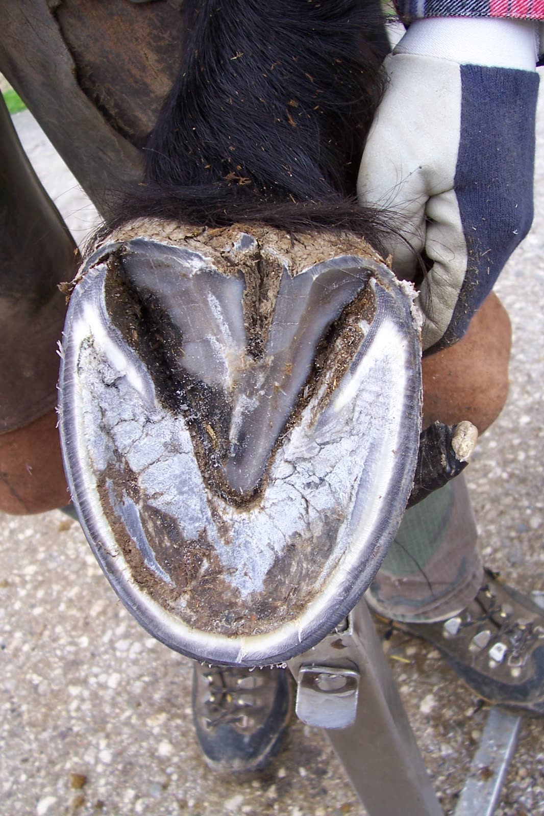
A horse's hoof is made of a hard, horny outer capsule and the living tissue and bone inside it. The outer rim you see when the horse is standing is the hoof wall (제벽). When the foot is picked up, a V-shaped structure called the frog (제차) sits in the middle of the underside. The wall, like a human fingernail, has no nerves or blood vessels and keeps growing. The frog is elastic like rubber; it absorbs shock and helps blood circulate through the foot. Knowing these two structures makes hoof trimming and shoeing much easier to understand.
The parts you see from outside: coronet and wall
The band at the top of the hoof, where the hairy skin meets the hoof, is the coronet (coronary band). The wall is produced here and grows downward. Wikipedia gives a growth rate of about 1 cm a month, and the University of Missouri Extension about three-eighths of an inch. Professor Yang Jae-hyuk's Equine Hoof Science puts it at 0.5–1.0 cm a month, depending on breed, use and nutrition. Serious injury to the coronet can stop the hoof from growing properly and leave a permanent defect.
The wall has different names by position: the toe at the front, the quarters on each side and the heels at the back. It is tallest at the toe and gets lower towards the back. It is 6–12 mm thick or more. In the front feet the wall is thickest at the toe, while in the hind feet it is more even. Above the heels are two rounded swellings called the bulbs.
The wall has three layers. The outer layer carries pigment, so a dark patch on the coronet skin shows as a vertical stripe below it. Inside that is an unpigmented layer that resists ground contact and supports the horse's weight. The innermost layer is the white line. The three layers grow down together as one mass. If they are not worn down by the ground, they stick out below the sole and become likely to break.
The parts you see from below: frog, sole and bars
With the foot lifted, the triangular frog sits in the middle. It reaches forward across about two-thirds of the sole and gets thicker towards the back, where it joins the bulbs. It has a central groove, and a deeper groove called the collateral groove runs along each side. It is dark grey and rubbery. Wikipedia describes the frog as the equivalent of the human fingertip.
The surface between the frog and the wall is the sole. It is not flat but concave, about 1–1.5 cm deep. According to Equine Hoof Science, the sole of a hind foot is more concave than that of a front foot. At the heels, the wall turns sharply inward and runs along the collateral grooves. These folds are the bars.
What the frog does: shock absorber and blood pump
The University of Missouri Extension lists the wall, bars and frog as the weight-bearing structures, and says the sole does not normally touch the ground. When the foot lands, the elastic frog usually touches down first. As it is pressed, it pushes up on the digital cushion, a flexible mass inside the heel, which flattens and spreads to the sides. The frog flattens too and pushes the bars apart, so the heels widen. When the foot is lifted, everything returns to its original shape.
This movement also helps circulation. When the foot is put down, the veins in the foot are squeezed and blood is pushed up the leg. When it is lifted, blood flows back in. For a horse, whose legs are thin and far from the heart, the hoof works as a pump. The Missouri publication notes that exercise increases blood flow in the foot and helps the hoof grow, while lack of exercise, dryness and poor nutrition slow growth.
Points for shoeing and trimming, and common mistakes
- The white line guides the farrier. It is a soft horny layer joining the wall and the sole, and it marks the border between the sensitive and insensitive parts of the hoof. Horseshoe nails enter at the outer edge of the white line and come out through the wall at an angle, about 15–20 mm above the ground surface.
- The white line is not the inner wall. Equine Hoof Science points out that many people confuse the two. The white band outside the white line is the inner layer of the wall, while the white line itself is the yellowish line further in.
- Removing too much sole is a common mistake. The sole is not built to carry weight from the ground. Its concave shape protects the sensitive structures above it and gives grip.
- The frog needs dry ground. A horse that always stands in wet conditions can develop thrush, a bacterial infection of the frog.
Laminitis, in which the hoof separates from the coffin bone (the last bone inside the hoof), is the most serious hoof disorder and causes severe lameness. If the hoof looks different from usual or the horse seems to be limping, do not judge it yourself; it is safest to consult a veterinarian and a farrier.